Computational Bioimaging for Living Organism Imaging

BANDUNG, itb.ac.id – Bioimaging may not be a familiar word for us. However, it is one of the fastest-advancing technologies in biology.
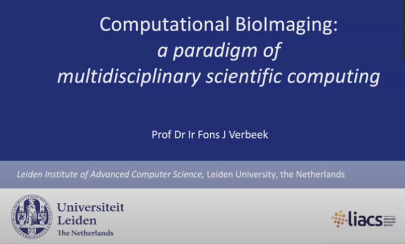
Through a seminar that was held by the Doctoral Program of the School of Natural Science and Technology ITB (SNST ITB) titled "Computational Bioimaging: A Paradigm of Multidisciplinary Scientific Computing" on Thursday (26/1/2022), Prof. Fons Verbeek from the University of Leiden, Netherlands, explained the bioimaging computational technology together with its implementation in the field of biology.
"Bioimaging is a form of visualization or biological object-imaging by minimizing all physical disturbances. In addition, bioimaging can gain information on the 3D structure of a specimen through external observation. This can include both subcellular and cellular observation from the scale of a tissue to a multicellular organism," said Prof. Verbeek.
One of the most well-known bioimaging applications is in examining cases that are invisible to the naked eye in the medical field or what is more commonly known as radiology. However, computational bioimaging can be implemented more broadly and involve a broader scientific field.
Computational bioimaging incorporates two well-known fields of information technology and data analysis: machine learning and deep learning. "The various features from machine learning used in computational bioimaging are first-order statistics, HU invariants, local binary patterns, discrete wavelet transform, and histogram of oriented gradients. Deep learning is utilized to prevent data overfitting and increase the tuning accuracy using data augmentation, transfer learning, cross-validation, and hard voting," explained Prof. Verbeek.
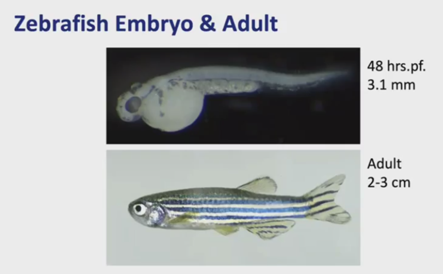
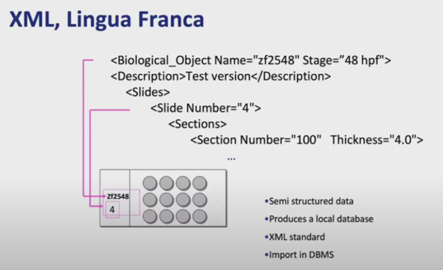
Some examples of bioimaging presented in the seminar were imaging a zebrafish embryo and a 3 cm-sized fish. The first step in obtaining the imaging is inserting the object on a petri dish into an image acquisition station section equipped with data processing software. The software that is used is written in an XML programming language with the following code sequence:
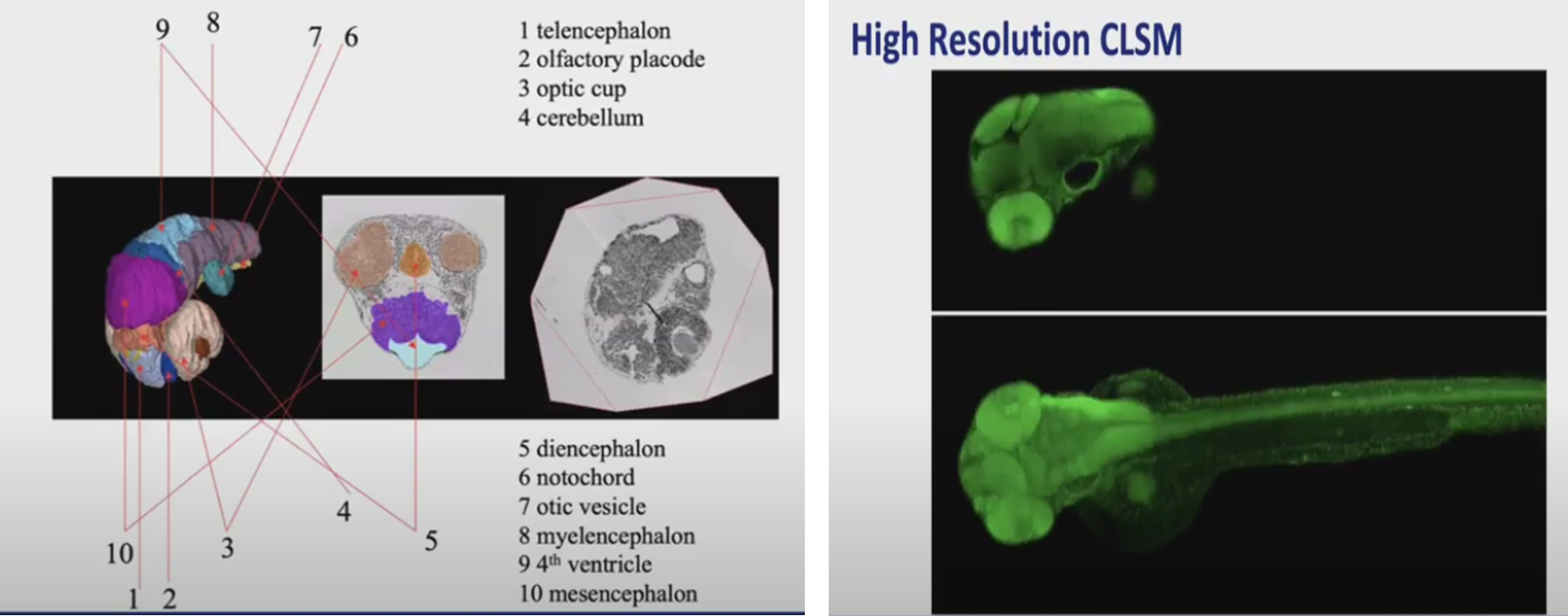
Then, after the object is registered, imaging can be obtained. This computational bioimaging allows various parts of the object to be observed in much clearer detail. Not only that, the resolution that is produced is very high (3000x2196). Moreover, imaging in the form of high-resolution CLSM and x-ray can also be obtained.
Reporter: Yoel Enrico Meiliano (Food Engineering, 2020)
Translator: Favian Aldilla R (Civil Engineering, 2019)

scan for download